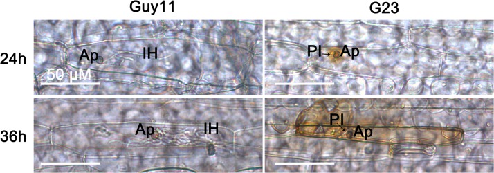
Fig 7. Detection of H2O2 accumulation at infected barley cells.

Detached barley leaf were inoculated with the tested strains, stained with DAB at 24 and 36 hpi, and observed under the light microscope. The cells infected by MoSFA1 deletion mutant were strongly stained with DAB, indicating high H2O2 accumulation at the penetration site. Bar is 50 μm. Ap, appressorium; PI, primary infectious hyphae; IH, secondary infectious hyphae.
