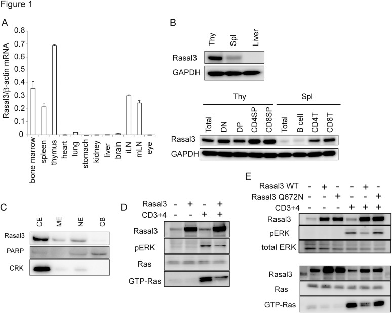
Fig 1. Expression, localization and function of Rasal3.
(A) Expression of Rasal3 mRNA in various organs from C57L/6 mice. Rasal3 mRNA expression was determined by quantitative RT-PCR and normalized relative to β-actin mRNA. (B) Expression of Rasal3 protein in the thymus, spleen, and liver (upper) and sorted thymic or splenic T cell or B cell subpopulations (lower). (C) Subcellular localization of Rasal3 protein in thymocytes. CE, ME, NE, and CB indicate cytoplasmic extract, membrane extract, nuclear extract, and chromatin bound, respectively. (D) GAP activity of Rasal3. DPK cells overexpressing Rasal3 were subjected to a Ras-GTP pull-down assay followed by Western blot analysis. (E) The Q672N mutant Rasal3 could not suppress the increase in GTP-Ras and ERK activation. All data are representative of more than three independent experiments.

