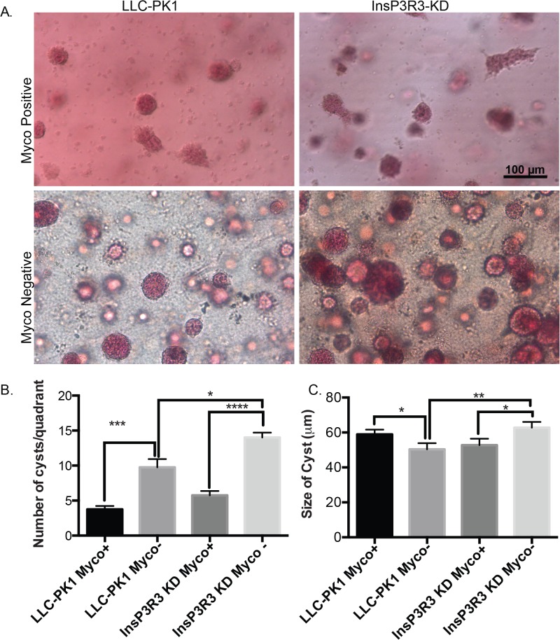
Fig 4. Cyst number is increased in 3D tissues without mycoplasma contamination.
(A) Mycoplasma positive and mycoplasma-negative cells were grown in 3D hydrogels for 2 weeks at which time the tissues were fixed and stained with Carmine for whole mount imaging. Note that cysts are more numerous in the mycoplasma negative tissues. Scale bars = 100 μm. (B) Quantified number of cysts across 4 different quadrants. (C) The diameter of the cysts is altered upon treatment of mycoplasma contamination. Data represents the average diameter and the error bars represent the standard error of the mean. * represents p>0.05, ** p>0.01, ***p>0.001, ****p>0.0001

