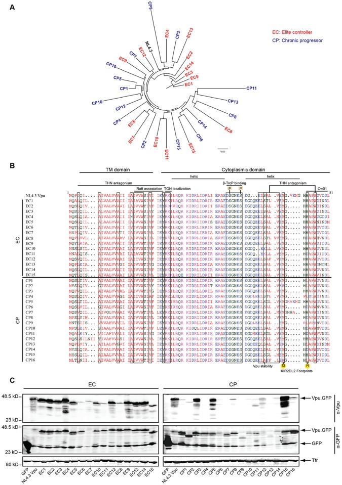
Fig 1. Maximum-likelihood phylogenetic tree, sequences alignment, expression and detection of plasma HIV RNA-derived Vpu clonal alleles.
A) Maximum-likelihood phylogenetic tree of HIV-1 Vpu alleles. EC-derived Vpus in red, CP-derived Vpus in blue, and control strain NL4.3 in black. B) Amino acid sequence alignment of the Vpu proteins analyzed generated using Clustal Omega (EMBL-EBI). HIV-1 NL4.3 Vpu on the top serves as reference sequence. Boxes indicate the position of functionally relevant residues and motifs. Different colors present the properties of the amino acid: red for small and aromatic, blue for acidic, green for residues with hydroxyl or sulfhydryl groups and magenta for positively charged residues. C: Expression and detection of Vpu alleles. Lysates of A3.01 cells transfected with Vpu.GFP expression plasmids were analyzed by Western blotting using antibodies against Vpu, GFP, and transferrin receptor (Tfr).

