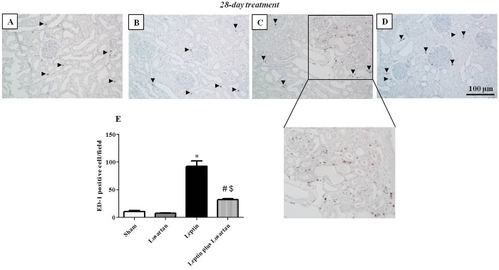
Fig 6. ED-1 staining.

Renal ED-1 immunostaining of the rats treated for 28 days: sham (A), losartan (B), leptin (C) and leptin plus losartan (D). ED-1-positive cell quantification (in cells/area) (E). Magnification of the leptin-treated rats ED-1 positive cell area. G = glomeruli; arrowheads = ED-1-positive cells; bar = 100 μm. *p<0.05 versus the sham; #p<0.05 versus the leptin-treated rats and $p<0.05 versus the losartan-treated. For the rats treated for 28 days: sham (n = 6); losartan (n = 6), leptin (n = 8); leptin plus losartan (n = 6).
