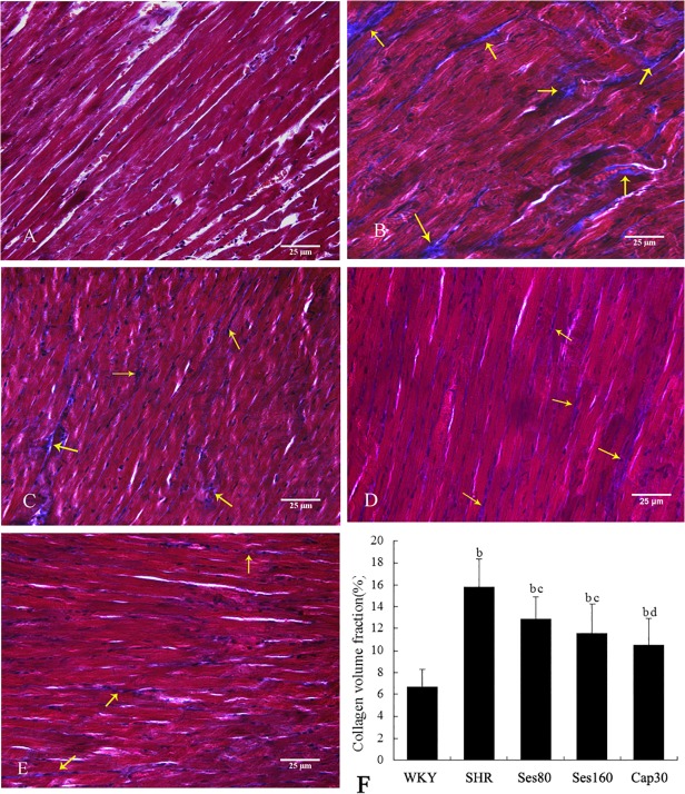
Fig 3. Effect of sesamin on left ventricular collagen deposition in rats.
Paraffin sections were subjected to Masson staining, followed by evaluation of collagen deposition with collagen volume fraction (CVF), the mean percentage of collagen area to the total area of each microscopic field. The CVF of each animal represents the mean of 6 randomly selected microscopic fields (400×). Data were presented as mean±SD (n = 7). A: WKY; B: SHR; C: Ses80; D: Ses160; E: Cap30; F: Quantitative analysis of collagen deposition (CVF). b P <0.01 vs. WKY group; c P <0.05, d P <0.01 vs. SHR group.

