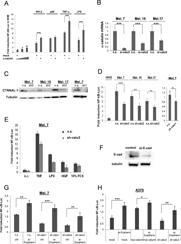
Fig 1. α-Catulin promotes NF-κB activation in human primary melanocytes and melanoma cells.
(A) A 5x NF-B-luc reporter gene (0.25μg) was co-transfected into melanocytes together with different concentrations of α-catulin (1 or 1.5 μg) with or without IKK-β(0.5μg) or p65 (0.5μg). 24 h later cells were non-stimulated or stimulated with TNF-α or LPS. Luciferase levels were normalized for a co-transfected RFP control (0.25μg). (B) Mel.7, Mel.15 and Mel.17 cells were stable infected with lentiviral particles containing a vector-based mirRNA construct directed against α-catulin (sh-catu 1 or sh-catu2; Materials and Methods), and α-catulin mRNA levels were analyzed by real-time PCR. (C) Mel.7, Mel.15 and Mel.17 cells were analyzed by Western blot with antibodies against α-catulin. (D) A NF-κB-luc reporter gene was tranfected into melanocytes and different melanoma cells containing stable integrated α-catulin mirRNA constructs (α-catulin-knockdown), and luciferase values were determined 24 h later and normalized for co-transfected JRED values. (E) Melanoma 7 cells as in (D) except that the cells were stimulated with TNFα, LPS, HGF and 10% FCS for further 8 h. (F) Mel.7 cells (n.s., sh-catu1/2) were transfected with NF-κB reporter plasmid and with or without si-RNA construct directed against E-cadherin and luciferase values were determined. (G) NF-κB-luc reporter assay with A375 melanoma cells transfected with mock, myc-α-catulin or sh-catu2 plasmids together with or without E-cadherin si-RNA. *Indicates P>0.005, **P>0.001, ***P>0.0001, Student´s t test.

