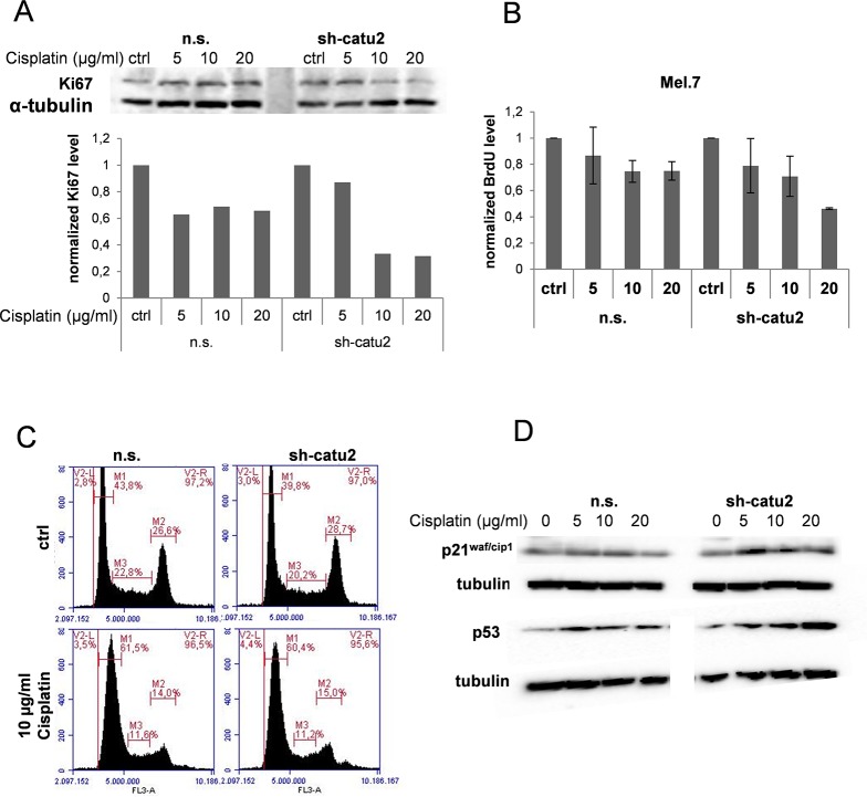
Fig 5. Cell proliferation is reduced in a dose- and time dependent manner in cisplatin treated melanoma cells when α-catulin is knocked down.
(A) Stable infected Mel.7 cells (n.s., sh-catu2) were treated with 0, 5, 10 and 20 μg/ml cisplatin for 24 h and analyzed by Western blot with antibody against Ki67. α-Tubulin was used as a loading control and quantification was performed with BioRad software. (B) Mel.7 cells (n.s., sh-catu2) were treated with 20, 10, 5, or 0 μg/ml cisplatin for 48 h and BrdU assay was performed. Therefore, cells were stained with BrdU solution and antibodies against BrdU and HRP conjugated secondary antibody was detected at 450 nm using a multiplate reader. (C) Mel.7 cells (n.s., sh-catu2) were treated with 0 or 10 μg/ml cisplatin for 18 hours, fixed, stained with propidium-iodide solution and analysed for cell cycle distribution using flow cytometry. (D) Cells as in (C) were analysed using western blot with antibodies against p21cip/waf and p53.

