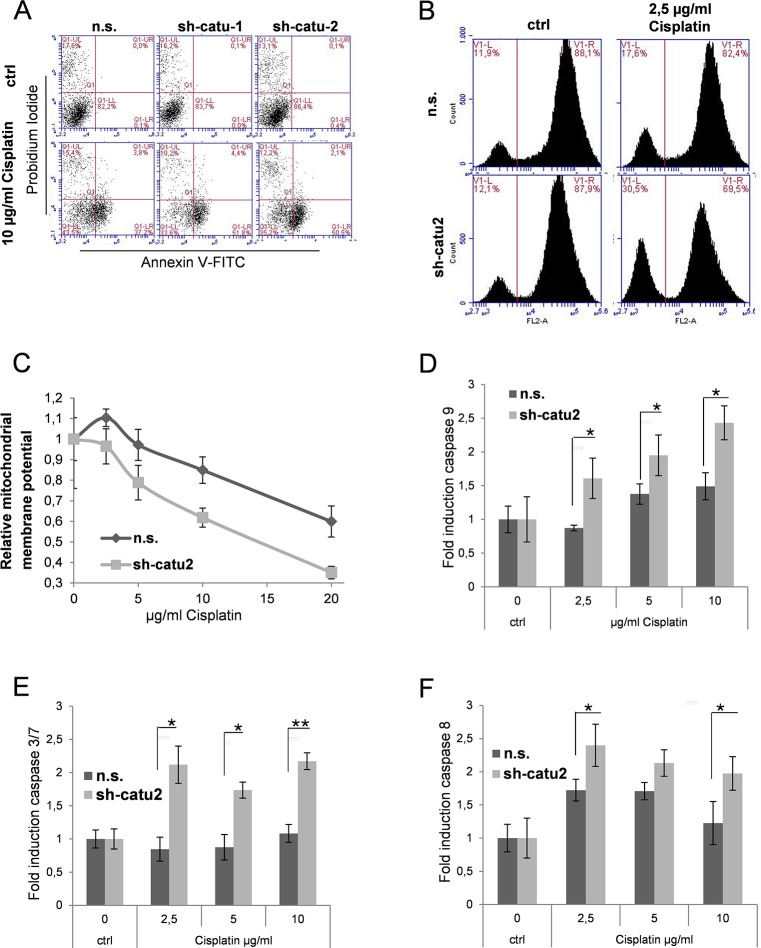
Fig 6. α-Catulin knockdown enhances apoptosis in cisplatin-treated melanoma cells.
(A) Stable infected Mel.7 cells (n.s., sh-catu1, sh-catu2) were treated with 0 and 10 μg/ml cisplatin for 48 h and stained with Annexin V and PI and analysed using flow cytometry (B) Stable infected Mel.7 cells (n.s., sh-catu2) were treated with 0 and 2.5μg/ml cisplatin for 6 h. For cytochrome c release assay, cells were treated with permeabilization buffer, fixed with formaldehyde, stained with antibody against cytochrome c and analyzed by Flow Cytometry. (C) Mel.7 cells (n.s., sh-catu2) were seeded in a 96 well plate and treated with 0, 2.5, 5, 10 or 20 μg/ml cisplatin for 6 h and mitochondrial membrane potential was determined using JC-1 assay (D) Mel.7 cells (n.s., sh-catu2) were treated with 0, 2.5, 5 or 10 μg/ml cisplatin for 6 h and analyzed for caspase 9 activity using caspase glo luminescence assay. (E) Cells as in (D) were analyzed for caspase 3/7 and (F) caspase 8.

