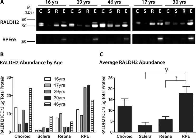
Fig 3. Western blot analysis and quantification of RALDH2 in postnatal human ocular tissues.
(A) Cytosol fractions from ocular tissues of donors aged 16–46 years were immunblotted with anti-RALDH2 or anti-RPE65. Top Panel: RALDH2 immunopositive bands (∼ 55–59 kDa) were present in the choroid (C), sclera (S), retina (R), and RPE (E) of postnatal human eyes. Bottom Panel: anti-RPE65 was used to identify the presence of RPE contamination in the retina, choroid, and sclera samples. 3 μg total protein/lane was loaded for each blot. (B) Relative abundance of RALDH2 was measured as the integrated optical density (IOD) per 3 μg total protein of RALDH2-immunopositive bands. (C) Average RALDH2 abundance (± s.e.m.) in ocular tissues of all donors presented in (B) (n = 5). *p < 0.05, **p < 0.01 (one-way ANOVA followed by Tukey-Kramer test for multiple comparisons).

