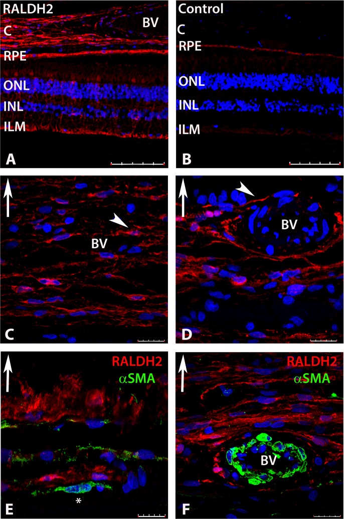
Fig 4. Confocal images of RALDH2 (red) and αSMA (green) expressing cells after immunolabeling with anti-RALDH2 and anti-αSMA.
(A) Low power magnification of ocular tissues demonstrating RALDH2 labeling in the retina, RPE, and choroid. (B) Negative control section (primary antibody pre-absorbed with recombinant RALDH2) demonstrating absence of labeling in the retina and choroid and slight auto-fluorescence in the RPE. (C, D) Choroid demonstrating RALDH2 labeling in extravascular cells closely associated with blood vessels and in slender cells throughout the stroma (arrowhead). (E, F) Double labeling of the choroid with anti-RALDH2 and anti-αSMA demonstrates RALDH2 positive cells are distinct from vascular and extravascular smooth muscle cells. Asterisk (*) indicates an αSMA-positive extravascular smooth muscle cell in (E). Upward arrow in (C-F) indicates orientation for the scleral side of the choroid tissue. Nuclei were counterstained with DAPI (blue). BV, blood vessel; C, choroid; RPE, retinal pigment epithelium; ONL, outer nuclear layer; INL, inner nuclear layer; ILM, inner limiting membrane. Scale bars = 100 μm in A, B; 20 μm in C-F.

