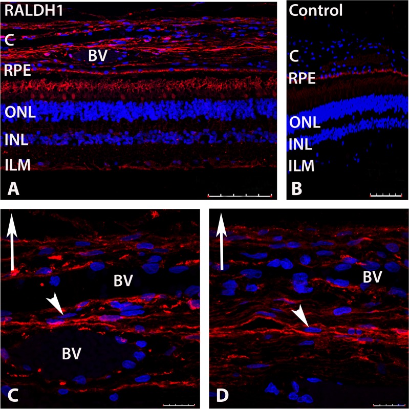
Fig 6. Confocal images of RALDH1 expressing cells in postnatal human ocular tissue after immunolabeling with an anti-RALDH1 antibody.
(A) Low power magnification of ocular tissues demonstrating RALDH1 labeling (red) in the retina and choroid. (B) Negative control slide (non-immune IgG used in place of primary antibody) demonstrating no labeling in the retina and choroid and auto-fluorescence in the RPE. (C, D) Choroid sections demonstrating RALDH1 labeling in extravascular cells throughout the choroidal stroma (arrowhead). Upward arrow in (C-D) indicates orientation for the scleral side of the choroid. Nuclei were counterstained with DAPI (blue). BV, blood vessel; C, choroid; RPE, retinal pigment epithelium; ONL, outer nuclear layer; INL, inner nuclear layer; ILM, inner limiting membrane. Scale bars = 100 μm in A, B; 20 μm in C, D.

