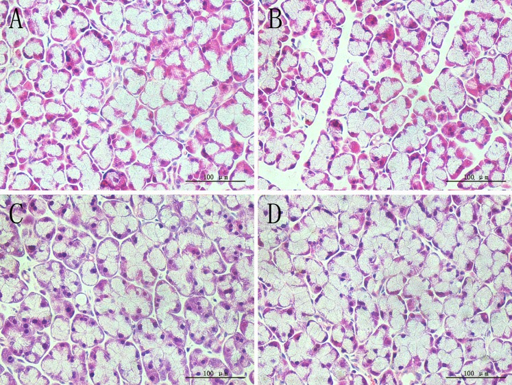
Fig 1. H&E staining of lacrimal gland.
A, SHAM; B, OVX; C, OVX + E; D, OVX + iCR. All scale bars represent 100 μm (magnification ×400). Compared with the SHAM group, the OVX group showed a sparse acinar cells, and the intercellular space was increased, suggested that cell atrophy was present, while estradiol valerate and iCR treatment can protected the glands from ovariectomy-induced cell atrophy to a certain extent.

