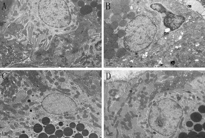
Fig 9. The GCT in the submandibular gland.
A, SHAM; B, OVX; C, OVX + E; D, OVX + iCR. Scale bars represent 1 μm. In the SHAM group, the cell plasma was filled with a high density of secretory granules, and the nucleus was located on the basolateral side of the cell with normal chromatin morphology, and mitochondria were intact. In the OVX group, many of the mitochondria were swollen, with double membrane structure damage and crest fracture, and “balloon-like” mitochondria were common. The condensed chromatin morphology was inside the cells, probably resulted- from the mutual ingestion of apoptotic cells. The color density of the secretory granules was reduced, with a gray appearance. In the OVX+ E and OVX+ iCR groups, most of the mitochondrial structure was normal, the distribution of chromatin was uniform, and the density of black secretory granules was similar to that of the SHAM group.

