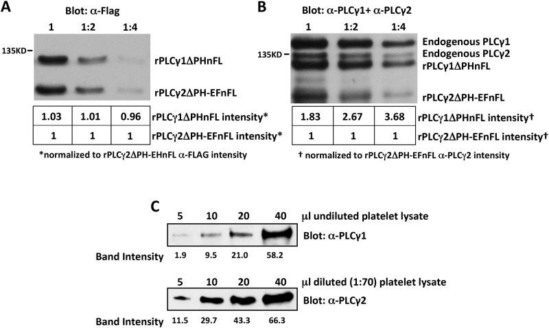
Fig 2. Quantification of relative levels of PLCγ1 and PLCγ2 in mouse platelets.
(A and B) Lysates were prepared from COS-7 cells transfected with a plasmid encoding a FLAG-tagged form of either PLCγ1 in which the pleckstrin homology domain was deleted (rPLCγ1ΔPHnFL) or PLCγ2 in which the PH domain and EF hands were deleted (rPLCγ2ΔPH-EFnFL) and mixed. (A) Serial two-fold dilutions of the mixed COS-7 lysates were subjected to Western blot analysis with antibodies specific for the FLAG tag. Numbers under each lane indicate the densities of the rPLCγ1ΔPHnFL and rPLCγ2ΔPH-EFnFL bands relative to that of the rPLCγ2ΔPH-EFnFL band, which was assigned an arbitrary value of 1. These data demonstrate that rPLCγ1ΔPHnFL and rPLCγ2ΔPH-EFnFL proteins were equally loaded in each lane. (B) The same serial dilutions of mixed COS-7 cell lysates were subjected to Western blot analysis with a mixture of PLCγ1- and PLCγ2-specific antibodies. Note that endogenous PLCγ1 and PLCγ2 are also detected by these antibodies. Numbers under each lane indicate the density of the rPLCγ1ΔPHnFL and rPLCγ2ΔPH-EFnFL bands relative to that of the rPLCγ2ΔPH-EFnFL band, which was assigned an arbitrary value of 1. These data demonstrate that the anti-PLCγ1 antibody recognizes PLCγ1 about three times better than the anti-PLCγ2 antibody recognizes PLCγ2. (C) Increasing amounts of undiluted (top) or 1:70 diluted (bottom) highly purified mouse platelet lysate were subjected to Western blot analysis with antibodies specific for PLCγ1 (top) or PLCγ2 (bottom). Numbers under each lane indicate the density of each band. Note that approximately 140X more platelet lysate was required to achieve a PLCγ1 band intensity equivalent to that of PLCγ2. Together with the finding that anti-PLCγ1 recognizes PLCγ1 approximately 3X better than anti-PLCγ2 recognizes PLCγ2, we conclude that mouse platelets have ~400X less PLCγ1 than PLCγ2.

