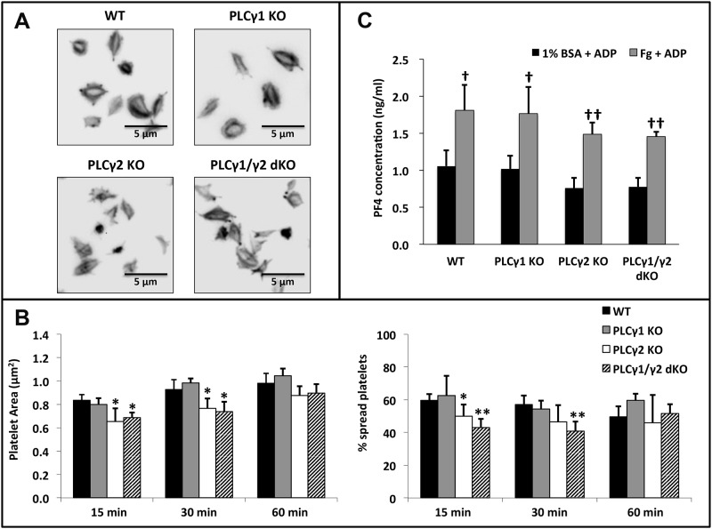
Fig 6. Effect of PLCγ1 or/and PLCγ2 deficiency on platelet spreading and granule secretion on immobilized Fg in the presence of ADP.
Washed platelets from WT, PLCγ1-deficient (PLCγ1 KO), PLCγ2-deficient (PLCγ2 KO) and PLCγ1/γ2 double-deficient (PLCγ1/γ2 dKO) mice were allowed to spread on Fg (3 μg/ml) in the presence of 20 μM ADP for up to 60 minutes at 37°C. (A) Platelets were fixed, permeabilized, and stained for F-actin using TRITC-Phalloidin. (B) Quantitative analysis of mouse platelets spread on immobilized Fg. Platelet spreading was quantified using Metamorph software (for each genotype, at least 200 platelets were analyzed), platelet spreading area (μm2) and percentage (%) are shown. Results are reported as mean ± S.D. from 3 independent experiments using 3 different groups of mice. (C) Assessment of granule secretion from spread platelets. Supernatants of platelets allowed to spread on fibrinogen in the presence of ADP for 60 minutes were collected and assayed for PF4 concentration by ELISA. Results are reported as mean ± S.D. from 3 independent experiments (*p < 0.05, **p < 0.001 for each genotype relative to wildtype; †p < 0.05, ††p < 0.005 for Fg + ADP relative to 1% BSA + ADP). Note that the failure of PLCγ2-deficient or PLCγ1/γ2 double-deficient platelets to spread or release granule contents on immobilized Fg was overcome by addition of exogenous ADP.

