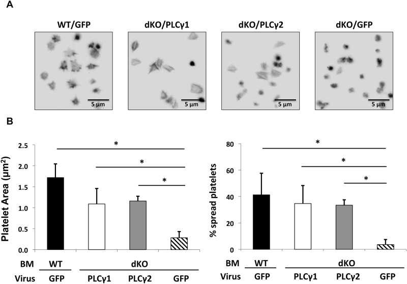
Fig 8. Restoration of αIIbβ3-mediated platelet spreading on Fg by enforced expression of PLCγ1 or PLCγ2 in PLCγ1/γ2 double-deficient platelets.
Lethally irradiated wild-type (WT) mice were reconstituted with WT bone marrow transfected with IRES-GFP retroviruses (WT/GFP) or with PLCγ1/γ2 double-deficient bone marrow transfected with IRES-GFP retroviruses (dKO/GFP), PLCγ1-IRES-GFP retroviruses (dKO/PLCγ1), or PLCγ2-IRES-GFP retroviruses (dKO/PLCγ2). Recipient mice were analyzed 8 weeks after reconstitution. (A) Washed non-sorted platelets from reconstituted recipients were allowed to spread on Fg (3 μg/ml) for 60 minutes at 37°C. Platelets were fixed, permeabilized, and stained for F-actin using TRITC-Phalloidin. (B) Quantitative analysis of mouse platelet spreading on immobilized Fg. Platelet spreading was quantified using Metamorph software (for each genotype, at least 300 platelets were analyzed), platelet spreading area (μm2) and percentage (%) are shown. Quantitative analysis was performed on all non-sorted platelets regardless of GFP positivity. Results are reported as mean ± S.D. from 2 independent experiments (*p < 0.05 for each genotype relative to dKO/GFP). Note that increased platelet spreading area and spreading percentage were observed in PLCγ1/γ2 double-deficient platelets that expressed PLCγ2 or PLCγ1 at levels normally achieved by PLCγ2.

