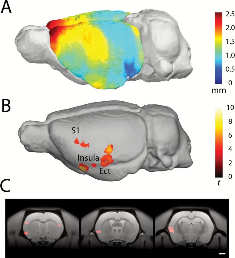
Figure 4.

Average voxel-based cortical thickness in rats treated with PCP. Scale bar in mm (A). Areas of significantly reduced cortical thickness in PCP-treated animals compared with saline-treated animals; the scale bar shows student’s t-score based on 14 degrees of freedom. Results surviving a cluster-extent correction for family-wise error p < 0.05 are shown (B). Coronal sections indicating reduced cortical thickness in the insular cortex, left somatosensory cortex (S1; C; panel 1), granular and dysgranular insular cortex (panel 2), granular, dysgranular insular cortex, and ectorhinal cortex (panel 3). Regions identified using Paxinos and Watson (1997).
