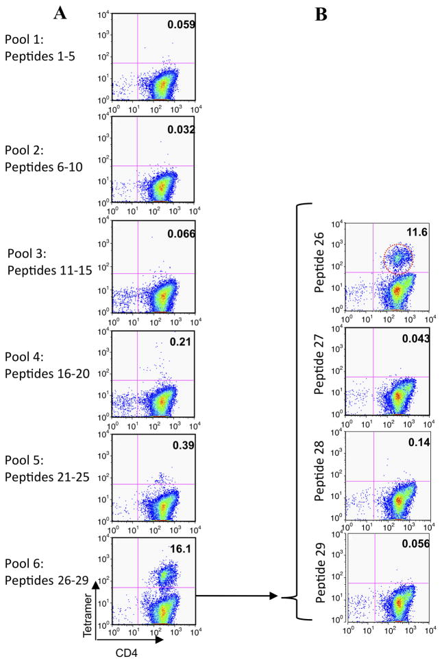
Figure 2.
Tetramer-guided epitope mapping identifies peptide 26 (p26) as a human leukocyte antigen (HLA)-DR1-restricted CD4T-cell epitope of Phl p 5b. A panel of overlapping 20-mer peptides spanning Phl p 5b was synthesised. Peptides were divided into pools and loaded onto PE-conjugated HLA-DR1 tetramers. Isolated CD4 T cells were cultured with adherent cells in the presence of the pooled peptides for 7 days before adding interleukin 2. Between days 12 and 14 the cells were stained with anti-CD4 and the pooled peptide tetramers (A). Positive tetramer staining was identified by flow cytometry. Individual peptide tetramers were generated and staining repeated (B). In this representative example, positive labelling is seen with HLA-DR1 tetramers loaded with p26 of Phl p 5b in patient URM031.

