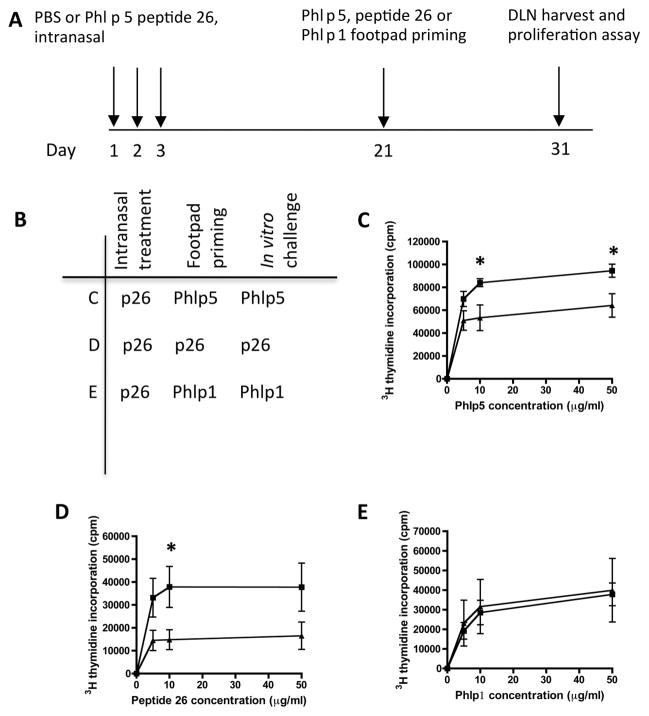
Figure 5.
Intranasal treatment with peptide 26 (p26) results in reduced T-cell proliferation to rPhl p 5b and p26. (A, B) Mice were treated intranasally with phosphate-buffered saline (PBS, filled squares) or p26 (filled triangles) at days 1, 2 and 3 and footpad primed at day 21 with (C) rPhl p 5 protein (each group, n=7), (D) p26 (each group, n=7) or (E) whole Phl p 1 protein (each group, n=5). Draining lymph nodes were harvested at day 31 and 3Hthymidine incorporation measured for protein or peptide as shown. An unpaired t test was used to determine significant differences between groups. Statistically significant differences were defined as p values of less than 0.05 (*p<0.05).

