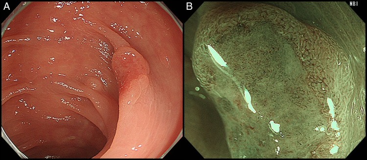
Figure 1.

Colonoscopic findings. (A) A 1 cm slightly reddish superficial elevated lesion with a shallow depression in the transverse colon. (B) Disrupted irregular microvessels and the absence of a surface pattern were observed under magnifying narrow-band imaging.
