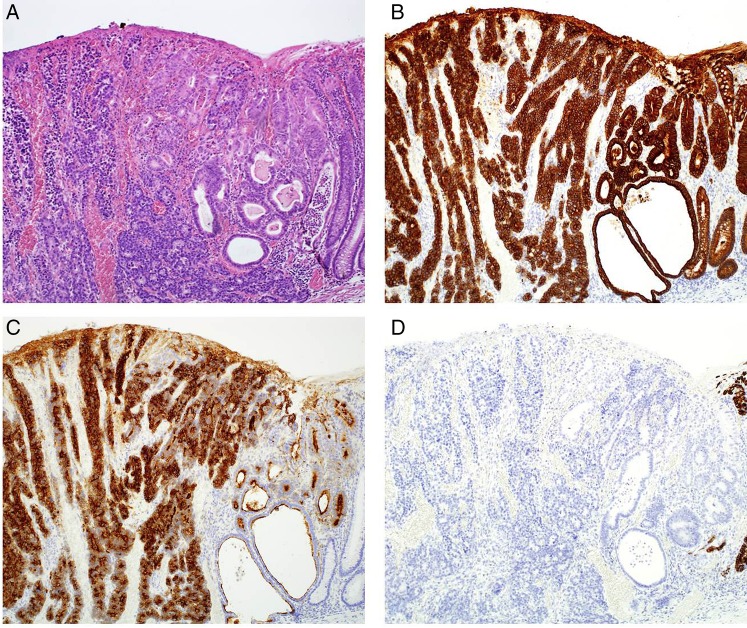
Figure 4.
Histological findings regarding the tumour. Two distinct components were observed. (A) H&E-stained tissue section. Neuroendocrine carcinoma and adenocarcinoma components can be seen. (B) Tissue section stained for cytokeratin AE1/AE3. Both components were positively stained. (C) Tissue section stained for CD10. The cytoplasm of the neuroendocrine carcinoma component and the brush border of the adenocarcinoma component were positively stained. (D) Tissue section stained for MUC2. Both components were not stained (magnification ×50).

