
Figure 4.
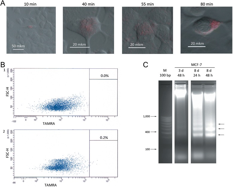
Analysis of hDNA internalization and apoptosis induction in MCF-7 cell line. А) Confocal imaging of TAMRA-labeled Alu DNA internalization by MCF-7 cells after 10, 40, 55 and 80 minutes of co-incubation. В) Internalization of TAMRA-labeled Alu DNA by MCF-7 cells (flow cytometry analysis): 1 – control cells without DNA; 2 – cells incubated with TAMRA-labeled DNA for 1 hour. С) Nucleosomal ladder formed by DNA isolated from MCF-7 cells which were cultured with exogenous hDNA and induced by TNFα to undergo apoptosis. 3 d, 8 d denote 3 and 8 days of co-incubation with DNA, accordingly; 24 h and 48 h indicate the duration of TNFα-induced apoptosis. Arrows point to the typical DNA bands found upon apoptotic DNA defragmentation.