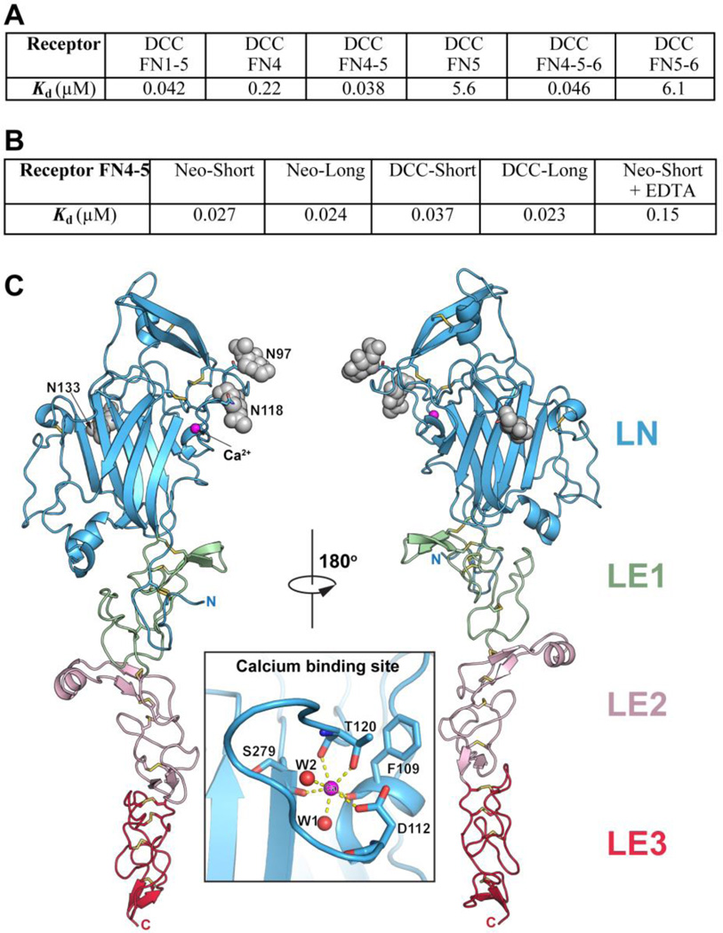
Fig. 2.
A) Binding of Netrin-1 (LN-LE1-LE2-LE3) to different DCC constructs documenting that the receptor FN4–FN5 region is necessary and sufficient for netrin binding. Kd, dissociation constant (in µM). B) Binding of Netrin-1 (LN-LE1-LE2-LE3) to the FN4–FN5 region of the different neogenin and DCC isoforms (see Fig. S4). To evaluate the role of the netrin-bound Ca++, 10 mM EDTA was added in one of the measurements. C) Structure of unbound Netrin-1. The individual netrin domains are labeled and colored in blue (LN), green (LE1), pink (LE2) and red (LE3). The glycosylation moieties at the three glycosylated Asn residues are shown as grey spheres. The N- and C- termini are labeled. The insert is a close-up view of the calcium-binding sire in the LN domain. The calcium ion is drawn in magenta and two bound water molecules – in red.

