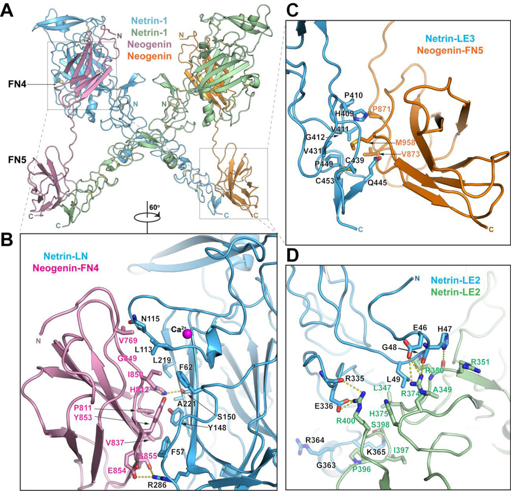
Fig. 3.
Structure of the Netrin-1/neogenin complex. A) Structure of the 2:2 Netrin-1/neogenin complex. The netrin molecules are in green and blue and the neogenin – in orange and magenta. The N- and C- termini of the molecules are labeled. B) Close-up view of the netrin-LN/neogenin-FN4 interface (Interface-1). Interacting residues and the netrin-bound calcium are labeled. C) Close-up view of the netrin-LE3/neogenin-FN5 interface (Interface-2). Interacting residues are labeled. D) Close-up view of the netrin-LE2/ netrin-LE2 interface (Interface-3). Interacting residues are labeled.

