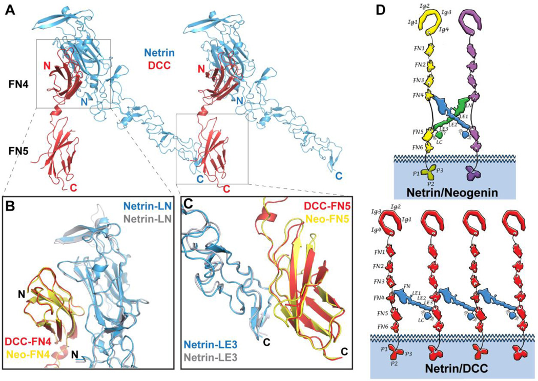
Fig. 4.
Structure of the Netrin-1/DCC complex. A) Structure of the Netrin-1/DCC complex. Netrin-1 is colored in blue, and neogenin in yellow and magenta. B) Superimposition of the LN-FN4 interaction site (Interface-1) in the Netrin-1/DCC (blue/yellow) and Netrin-1/neogenin (grey/pink) complexes. C) Superimposition of the LE3-FN5 interaction site (Interface-2) in the Netrin-1/DCC (blue/magenta) and Netrin-1/neogenin (grey/orange) complexes. D) Schematic drawing comparing the distinct Netrin-1/neogenin and Netrin-1/DCC signaling assemblies. The individual netrin and receptor domains are labeled. Ig, immunoglobulin; FN, fibronectin; LN, laminin-like N-terminal; LE, laminin-like EGF; LC, laminin-like C-terminal. Top, schematic representation if the 2:2 Netrin-1/neogenin complex. The netrin molecules are colored in blue and green, and the neogenin – in yellow and magenta. Bottom, schematic representation if the continuous Netrin-1/DCC assembly. The netrin molecules are colored in blue, and the DCC – in red. Interestingly, in both Netrin-1/DCC and Netrin-1/neogenin assemblies, the positively-charged netrin LC domain would be positioned towards the negatively-charged plasma membrane, thus potentially further stabilizing the signaling complexes at the neuronal surface.

