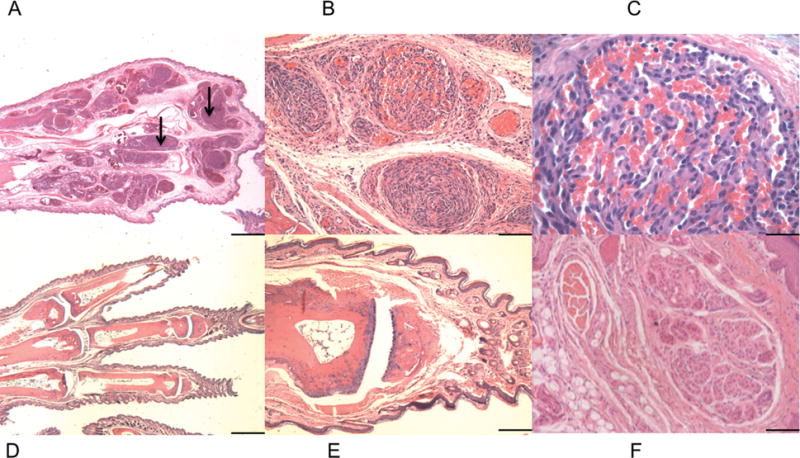
Figure 2.

Tsc1cc DARPP Cre mouse paw pathology. A-F) Hematoxylin and Eosin staining of paw lesions show extensive clusters of aberrant vascular cells and multiple hemangiosarcomas. A–C) Paw from 8-week-old mutant mouse, at 2× (A), 20× (B), and 60× (C) magnification. Black arrows indicate paw hemangiosarcoma tissue. D–F) Paw from 8 week old wild type mouse at 2× (D), 10× (E), and 20× (F). 2× scale bar = 1 mm, 10× scale bar = 200 um, 20× scale bar = 100 um, 60× scale bar = 33 um.
