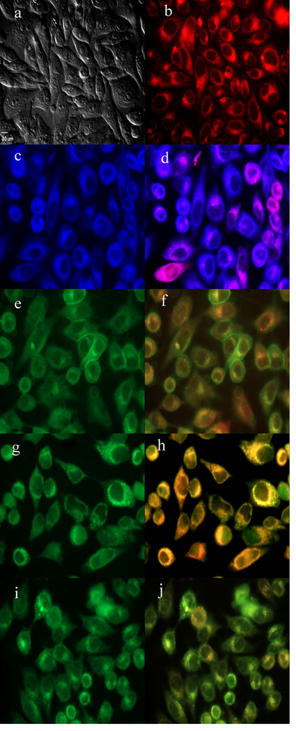
Figure 6.
Subcellular localization of BODIPY 2a in HEp2 cells at 10 µM for 6 h. a) Phase contrast, b) 2a fluorescence, c) ER tracker blue/white fluorescence, e) MitoTracker green fluorescence, g) BODIPY Ceramide, i) LysoSensor green fluorescence, and d), f), h), and j) overlays of organelle tracers with the BODIPY fluorescence. Scale bar: 10 µm.

