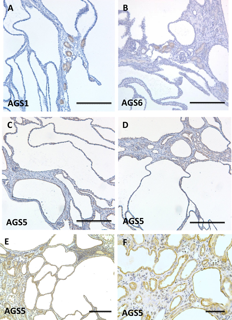
Figure 5.
Localization of Group I and II AGS3 proteins in polycystic kidney disease. Immunohistochemical staining was performed in mouse Pkd1V/V (A–D) and rat (E–F) PCK kidney models of polycystic kidney disease (PKD). AGS1, AGS5, and AGS6 were immunostained in kidneys sectioned from the Pkd1V/V mouse at post-natal day 19. In the PCK rat kidneys at 26 weeks of age, AGS5 immunostaining was performed and shown in (E) and (F). Scale bars = 200 µm (A–E), 50 µm (F).

