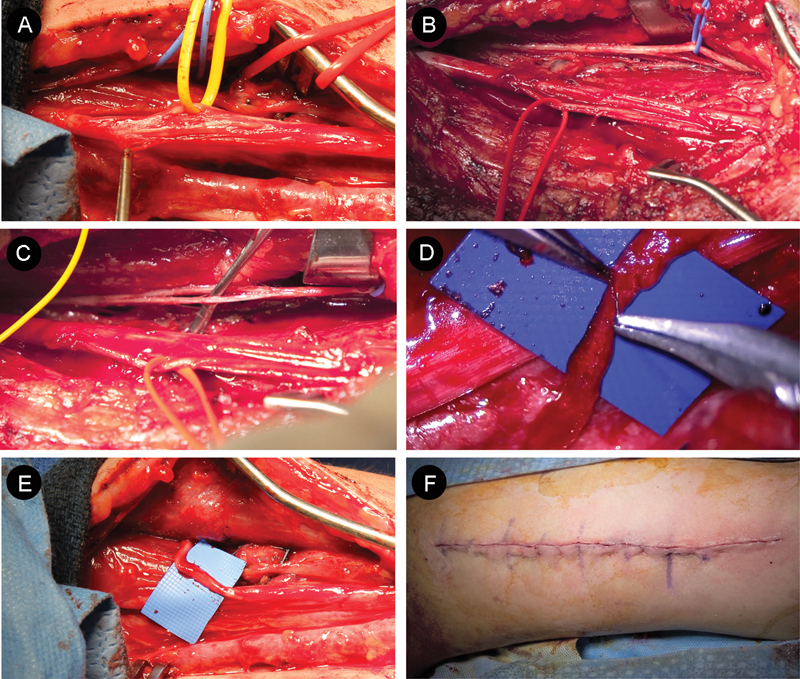
Fig. 3.

Transfer of brachialis nerve to anterior interosseous nerve (AIN) for C7 spinal cord injury: microdissection and anastomosis. (A) Nerve to the pronator teres fascicles (yellow loop) is located in the anterior portion of the median nerve. (B) AIN (red) and brachial nerve (blue) are identified and marked with vessel loops. (C) The brachialis branch is dissected distally to allow a tension-free repair. (D) Using a surgical microscope, the brachial nerve donor and AIN recipient are anastomosed, end-to-end. (E) Care is taken to ensure a tension-free repair. (F) The wound is closed in layers, a dressing is applied, and a sling is used to keep the elbow in a flexed and supinated position.
