
Figure 4.
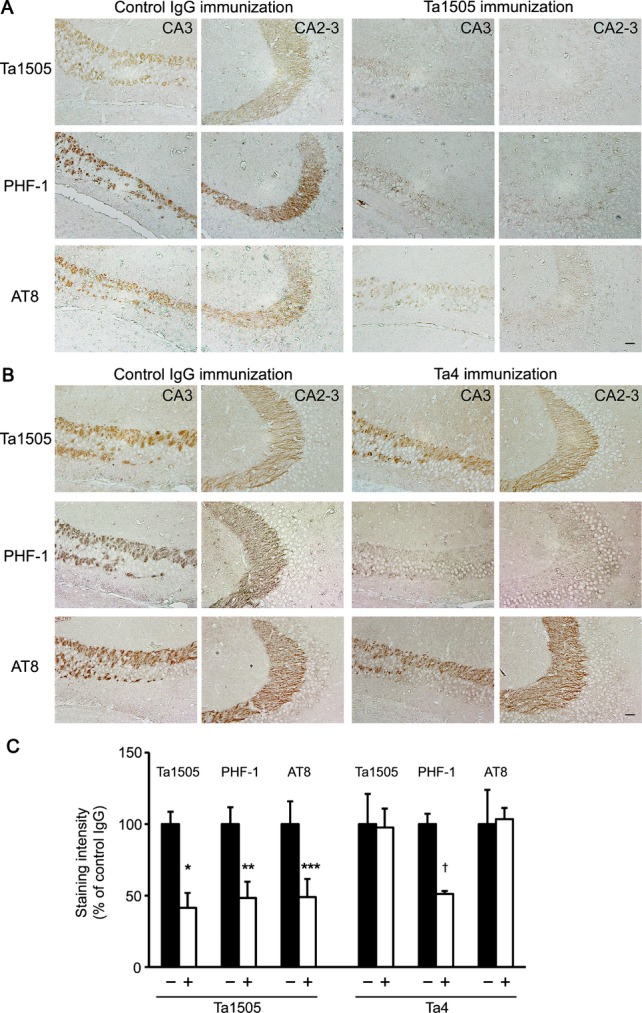
Effects of monoclonal antibodies on hyperphosphorylated tau in tauopathy mice based on immunohistochemistry. (A and B) Brain sections from 15-month-old antibody-treated tau784 mice were stained with Ta1505, PHF-1, and AT8 antibodies. Ta1505-treated mice showed an apparent reduction of hyperphosphorylated tau in hippocampal mossy fibers (A). In contrast, Ta4-treated mice exhibited reduced PHF-1-staining but not Ta1505- or AT8-staining (B). CA3 and CA2-3, hippocampal CA3 and CA2-3 regions. Scale bar, 30 μm. (C) Ta1505-, PHF-1-, and AT8-positive areas in each photograph of the hippocampal CA2-3 region were quantified using NIH ImageJ software. n = 5 for each group, except for control IgG in the Ta4 experiment where n = 3. *P = 0.0026, **P = 0.0133, ***P = 0.0364, and †P = 0.0002 versus control IgG.