
Figure 7.
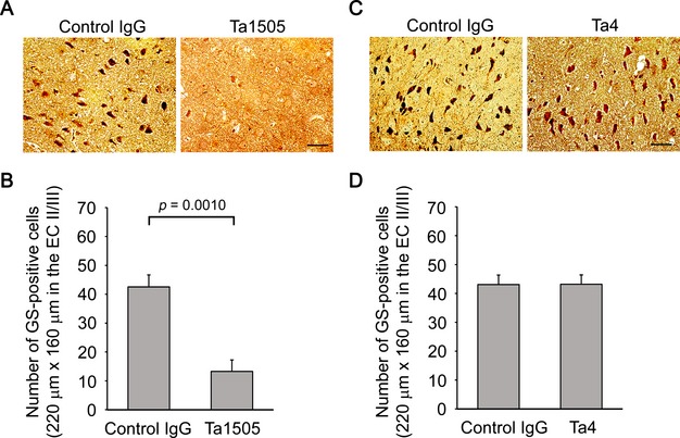
Effects of monoclonal antibodies on NFT formation in tauopathy mice. (A and C) Brain sections from 15-month-old antibody-treated tau784 mice were examined for NFTs by Gallyas silver staining. Ta1505-treated mice showed apparently reduced levels of NFTs (A), whereas Ta4-treated mice exhibited no changes (C). All images were taken from the EC-II/III region. Scale bar, 30 μm. (B and D) Gallyas silver positive cells in an area (220 × 160 μm) of the EC-II/III region were counted. (B) n = 5 for each group. (D) n = 4 for each group. NFT, neurofibrillary tangle; EC, entorhinal cortex.