
Figure 7.
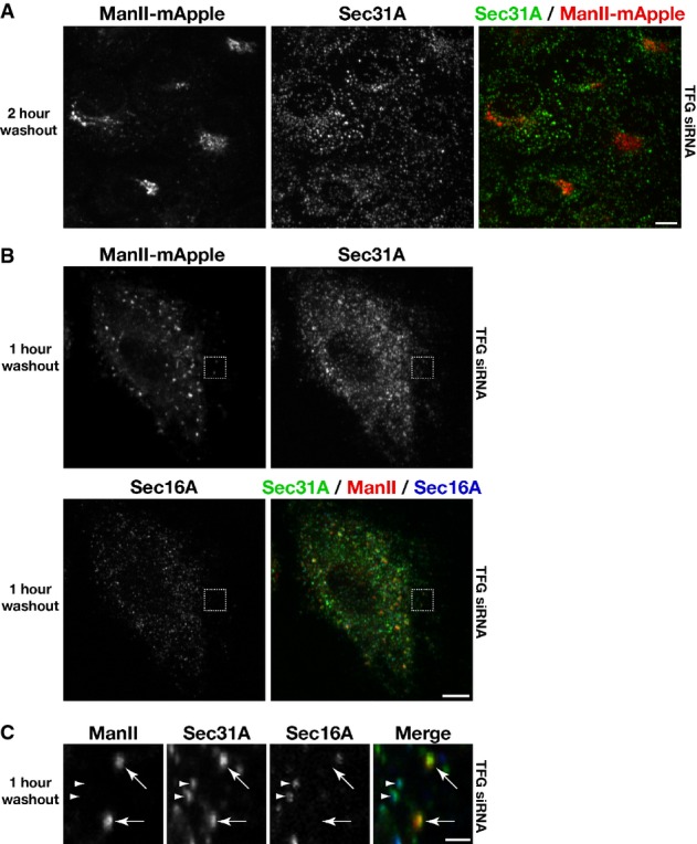
TFG depletion causes the accumulation of cargo-laden COPII transport carriers throughout cells
- Human RPE-1 cells stably expressing low levels of mannosidase II-mApple were transfected with a TFG siRNA for 60 h. Cells were subsequently treated with brefeldin A (BFA) for 1 h, followed by a wash into fresh media and further incubation for 2 h in the absence of BFA, prior to fixation. Images shown are projections of 3D data sets (4 μm in z). Merged images with Sec31A in green and ManII in red are shown (representative of at least 15 cells analyzed). Scale bar, 5 μm.
- Human RPE-1 cells stably expressing low levels of mannosidase II-mApple were transfected with a TFG siRNA for 60 h. Cells were subsequently treated with BFA for 1 h, followed by a wash into fresh media and further incubation for 1 h in the absence of BFA, prior to fixation and staining using α-Sec16A and α-Sec31A antibodies. Images shown are projections of 3D data sets (4 μm in z). Merged images with Sec31A (green), ManII (red), and Sec16A (blue) are shown (representative of at least 15 cells analyzed). Scale bar, 5 μm.
- Higher magnification views of the indicated regions in (B, boxed) are shown. Arrows highlight COPII-positive transport carriers that contain the cargo ManII, which are not juxtaposed to Sec16A-labeled sites on the ER. Additionally, arrowheads point out distinct foci in which COPII continues to associate with Sec16A-labeled sites, indicating that COPII vesicle formation continues in the absence of TFG. Scale bar, 1 μm.