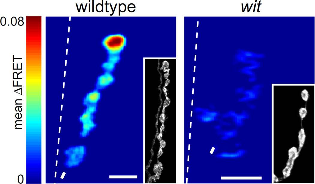
Figure 1.
Synaptic transmission is decreased in wit mutants. Mean SynapCam ΔFRET for wildtype and witHA2/witHA3 at 1.5 mM Ca2+ all on the same pseudocolor scale for ΔFRET (scale: 0–0.08). Throughout the paper, asterisks mark distal ends, dashed lines are borders between muscle 6 and 7, and arrows are where axons enter muscle or branchpoints. Insets: HRP stain showing the morphology of the NMJ. Scale bars = 5 µm throughout paper.

