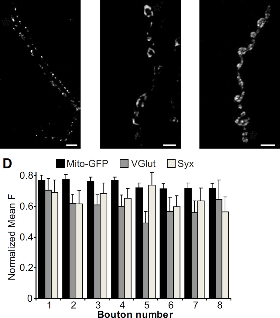
Figure 7.
There are no axonal gradients of mitochondria, synaptic vesicles, or a presynaptic T-SNARE. (A) NMJ expressing a GFP-tagged cytochrome C in motor neurons using D42-Gal4 to visualize mitochondria (D42-Gal4, UAS-mito-GFP/TM6). The asterisk indicates the distal bouton in a chain. (B–C) Wildtype NMJs stained for VGlut (B) and Syntaxin (C). (D) Quantification of mean fluorescence versus bouton position (Mito-GFP: n=35; VGlut: n=13; Syx: n=10).

