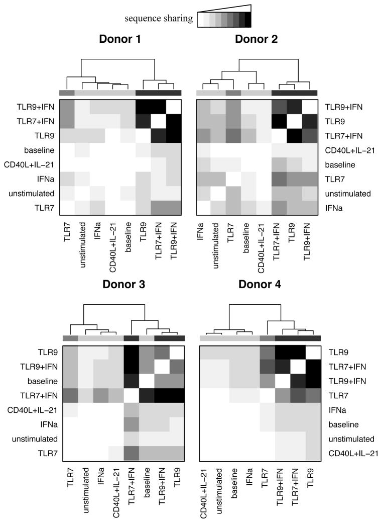
Figure 5. Deep sequencing of the IgH locus shows convergence of BCRs following TLR stimulation.
TLR9 and TLR/IFN activated B cells of each donor demonstrated increased CDR3 amino acid sequence sharing following stimulation, while two similarly showed convergence following TLR7 stimulation (sequence sharing as indicated.) To control for differing sequencing depth, repertoire subsets of equal size were examined pair-wise for all samples from each donor, counting overlapping sequences. These counts were then clustered based on complete clustering of Euclidean distances, with dendrogram shading added for ease of viewing. The greatest extent of overlap represents 0.5%, 3.9%, 3.3%, and 1.75% sharing of sequences for donors 1 through 4. Data represent four independent donors, split across two independent sequencing runs, with subsampling performed 5 times.

