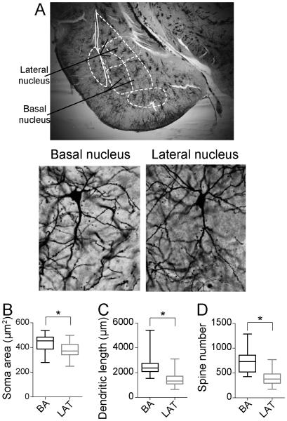
Figure 1. Morphological differences across nuclei in adolescent rats.
A) BA and LAT regions can be defined in Golgi-stained tissue with the aid of landmarks (top). Pyramidal-like neurons of the BA and LAT (bottom) were reconstructed after Golgi-Cox stain. The somatic area (B), average dendritic length (C) and average spine number (D) of principal neurons were significantly larger in the basal nucleus (BA) compared to the lateral nucleus (LAT) of adolescent rats. Average presented here as box plot with 5-95th percentile. * indicates p<0.05 in two-tailed t-test.

