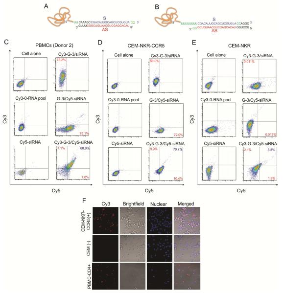
Figure 3. The design and binding affinity of aptamer-siRNA chimeras.
A, B) Schematic of CCR5 aptamer– siRNA chimeras. The region of the aptamer is responsible for binding to CCR5, and the siRNA is targeting TNPO3 gene. A linker (2 or 8 Us) between the aptamer and siRNA is indicated in green. Two versions, G-3-TNPO3 OVH chimera (A) and G-3-TNPO3 Blunt chimera (B), were designed. C, D, E) Cell surface binding of fluorescent dye –labeled RNAs was assessed by flow cytometry. The Cy3-labeled 0-RNA pool and Cy5-labeld siRNA were used as negative control. G-3-TNPO3 OVH chimera was chosen for binding affinity test with C) PBMC-CD4+ cells, D) CEM-NKr-CCR5 positive cells, E) CEM negative cells. The aptamer-sense strand and antisense strand of the chimera were labeled by Cy3 and Cy5 dye, respectively. And then they were annealed to form aptamer-siRNA chimera. F) Internalization analysis. The images were collected using 40×magnification as described previously.

