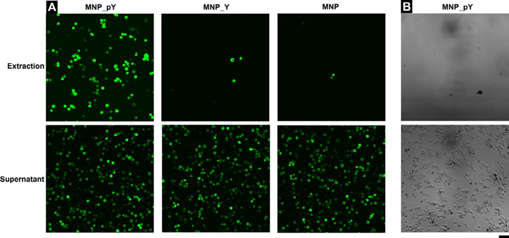
Figure 3.
(A) The fluorescent images (×20 dry objective lens) of the extraction and supernatant portions of cells after adding MNP_pY (Left), MNP_Y (Middle), and MNP (Right) to HeLa-GFP cells. (B) The bright field images (×20 dry objective lens) of the extraction and supernatant portions of cells after adding MNP_pY to HS-5 cells. Cells were incubated with the growth medium, Dulbecco’s Modified Eagle Medium (DMEM), containing 40 μg/mL nanoparticles for 4 hrs (top: the cells extracted by magnet; bottom: the cells remained in supernatant). The initial number of cells is 1.0×10⁶ per 6 cm culture dish. The scale bar is 100 μm.

