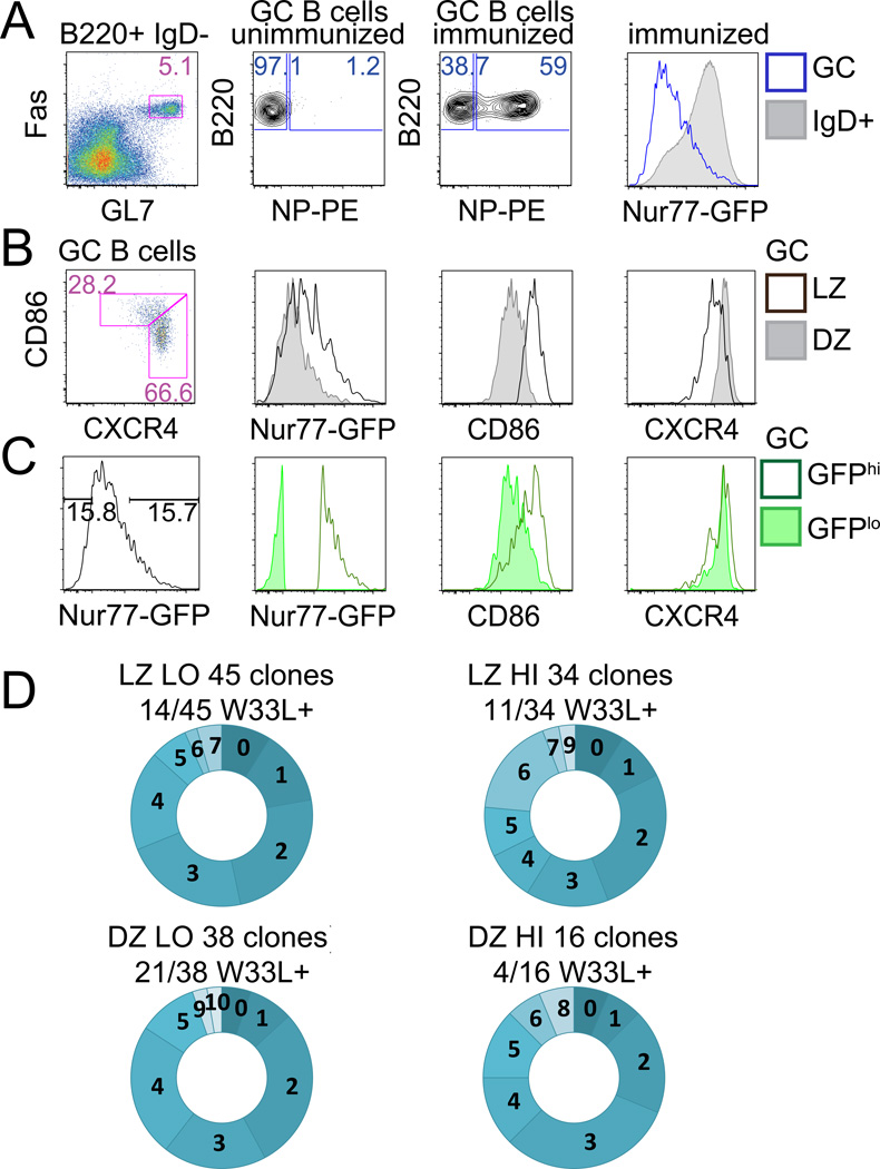
Figure 2. High Nur77-GFP-expressing GC B cells exhibit SHM.
(A–C) C57/Bl6 reporter splenocytes were stained d14 after NP-KLH immunization. (A) Plots represent gating to identify B220+IgDnegFashiGL7hi GC B cells (left hand panel), and NP-binding GC B cells (middle panels). Overlayed histograms (right hand panel) represent GFP expression from IgD+ (shaded histogram) and GC B cells (blue line). (B,C) Plots and histograms as described in Fig. 1E, F. Data are representative of 8 biological replicates.
(D) Vh186.2 heavy chains isolated from singly sorted GC B cells (see Fig. S1B for gating) from B6 reporter mice d10 after NP-KLH immunization were sequenced. Graphs represent number of mutations in individual clones (see Fig. S1C for mutation distribution). Numbers above graphs show proportion of clones with the W33L mutation.

