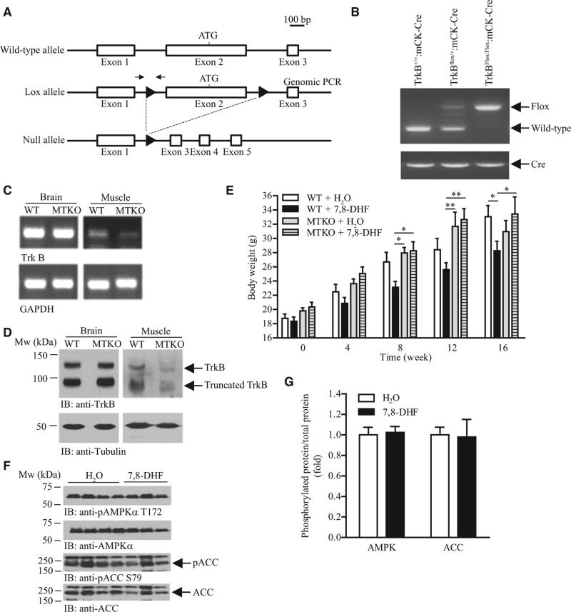
Figure 6. Muscle TrkB Is the Major Target of 7,8-DHF to Prevent Diet-Induced Obesity.
(A) A schematic representation of mouse TrkB deletion using loxP/Cre recombination. The location of loxP sites were marked as solid triangles. Locations of the primers used in the genomic PCR are indicated by the arrows.
(B) Deletion of TrkB in MTKO mice. Genomic DNA were isolated from the tail tip of female mice and used to perform PCR.
(C) Reduced TrkB expression in the skeletal muscle of MTKO mice. Total RNA was isolated from various tissues of female MTKO mice and used to generate cDNA. PCR was then performed to determine TrkB expression. Expression of GAPDH was also tested as control.
(D) MTKO mice have less TrkB protein in the skeletal muscle cell lysates were prepared from various tissues of female MTKO mice and the amount of TrkB proteins was examined using immunoblotting.
(E) Growth curve of 8-week-old female mice fed with HFD and 7,8-DHF in the drinking water (0.16 mg/ml) (*P < 0.05, **P < 0.01, two-way ANOVA, n = 7–10).
(F) Immunoblotting analysis of AMPK and ACC phosphorylation in muscle isolated from female MTKO mice that have been treated with 7,8-DHF for 16 weeks.
(G) Quantification of the protein phosphorylation shown in (F) (n = 3).
Results were presented as means ± SEM.

