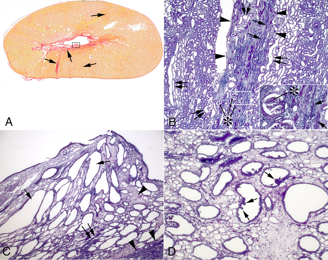
Figure 5.
Cortical and medullary changes in SWL-treated kidneys. Panel A show a transverse section of the upper pole of a treated kidney stained with Weigert’s hematoxylin and picro sirius red. Fibrosis extends from the renal capsule to the medulla (arrows) on both sides of the kidney. The largest cortical fibrotic band in panel A is shown in panel B (outlined in arrowheads) and shows sclerotic glomeruli (asterisks), atrophic tubular segments (arrows) and interstitial fibrosis—clearly seen in the panel B insert. Dilated tubule segments were found within fibrotic bands (double arrows) but more commonly in adjacent regions (double arrows). Panel C shows a damaged papilla outlined in a rectangle in panel A. Extensive interstitial fibrosis (arrowheads) surrounds dilated collecting ducts (arrows) and atrophic tubular segments (double arrow). A damaged papilla from a different kidney (panel D) shows a polypoid configuration (arrows) of the lining cells of several dilated inner medullary collecting ducts. Reduced from ×2 (A), ×300 (B), ×600 (B insert), ×1000 (C) and ×1500 (D).

