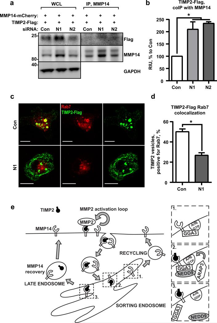
Figure 7. TIMP2/ MMP14 complex targets to Rab7 endosomes.
(a)WB analysis of WCL and IP with anti-MMP14 antibodies from siControl (Con) or siNEDD9 (N1, N2) MDA-MB-231 cells transfected with mCherry-MMP14 and Flag-tag fusion TIMP2; WB with anti-Flag and -MMP14 antibodies. (b) Quantification of WB as in (a) from three independent experiments; graphs are mean RIU values as % to siCon (assigned as 100%) ±S.E.M; one-way ANOVA with Dunnett's post-hoc analysis *p=0.0368 or 0.0216 for siCon/siN1 or N2; (c) Representative 3D reconstructed confocal images of cells as in (a) stained with anti-Rab7 (red) and anti-Flag antibodies (green); Scale bar is 10μm. (d) Quantification of colocalization of Rab7 and TIMP2 positive vesicles, mean % of TIMP2/Rab7 positive vesicles ±S.E.M; three independent experiments, 10 cells/treatment; two-tailed t-test:*p= 0.0003 for Con/N1. (e) Schematic representation of the model depicting action of NEDD9 on Afr6 through recruitment of ARAP3 to Arf6/GGA3/MMP14 complex.

