Figure 3.
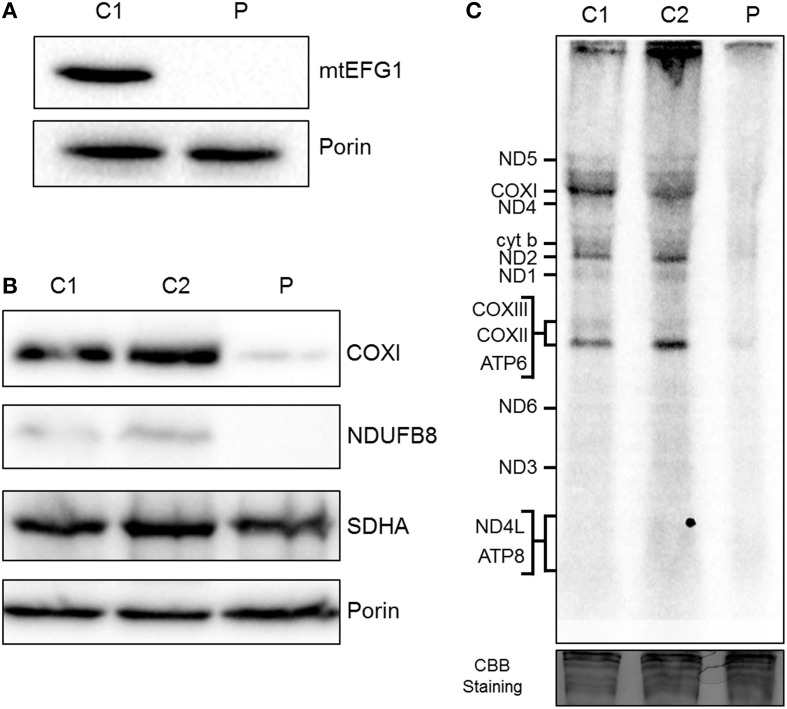
Biochemical assessment of patient fibroblasts. (A) Mitochondria isolated from control (C1) and patient (P) fibroblasts were subjected to SDS-PAGE and western blot analysis using an anti-mtEFG1 antibody. Anti-porin (VDAC1) antibody was used as a loading control. (B) Whole cell lysate from control (C1 and C2) and patient (P) primary fibroblasts were subjected to western blot analysis. Antibodies against COXI (mitochondrial encoded subunit of Complex IV), NDUFB8 (nuclear encoded subunit of Complex I), SDHA (nuclear encoded subunit of Complex II) and porin/VDAC1 (mitochondrial loading control). (C) Control (C1 and C2) and patient (P) fibroblasts were treated with emetine dihydrochloride to inhibit cytosolic translation and mitochondrial protein synthesis analyzed by [35S] met/cys incorporation (1 h followed by a 10 min chase). Cell lysate (50 μg) was separated through a 15% polyacrylamide gel. The gel was stained with Coomassie blue (CBB) to confirm equal loading. Post fixation and drying the signal was visualized by Typhoon FLA9500 PhosphorImaging. Signals were ascribed following established migration patterns (Chomyn, 1996).

