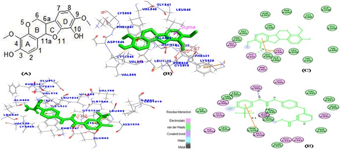
Figure 6.

The favorable binding portion of indigocarpan with lowest binding free energy in the ATP-binding site of VEGFR2 (PDB ID: 3VHE) as analyzed by molecular docking study. (a) 2D structure of Indigocarpan, (b) The three dimensional diagram displays the interaction of indigocarpan (the green stick) to the ATP binding site of VEGFR2 with the labeled amino acid residues CYS 919 and LYS 920 which significantly contributed to the binding. (c) The two dimensional diagram shows the interactions of indigocarpan to the amino acid residues. (d, e) denotes the binding mode of Sorafenib with VEGFR2. Similarly, colors of the residues indicate the forms of interactions as follows: van der Waals forces, green; polarity, magenta. Green arrow represents Hbonding with the amino acid main chain.
