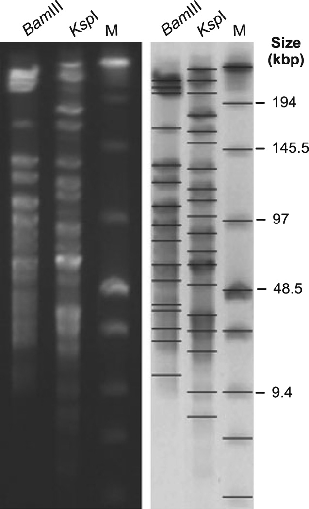
Figure 6.
Pulsed-field gel electrophoresis (PFGE) analysis of Helicobacter magdeburgensis. Chromosomal DNA was digested with the restriction endonucleases BamIII and KspI, respectively. Low Range PFGE Marker was used as the DNA size marker (M). BioNumerics software was used to identify bands and to determine band sizes. The values from the genome calculations are summarized in Table 2.

