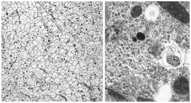
FIGURE 1.

Microscopic appearance of GSD III. Left, The hepatocytes are plant-like with clear cytoplasm and prominent cell membranes. The nuclei are small and uniform. Scattered glycogenated nuclei are seen. No cholestasis or significant inflammation is present (hematoxylin and eosin stain, original magnification ×20). Right, Hepatocyte with vast pools of particulate cytoplasmic glycogen. Rare lysosomes filled with glycogen are also seen (original magnification ×58,000).
