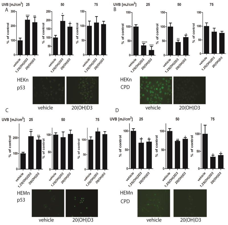
Figure 10.
Treatment of keratinocytes or melanocytes with 1,25(OH)2D3 or 20S(OH)D3 decreases CPD levels and increase p53 phosphorylation at Ser-15 following UVB exposure. Keratinocytes (A,B) and melanocytes (C,D) were treated with 100 nM 1,25(OH)2D3 or 20S(OH)D3 for 24 h prior to UVB exposure. Cells were exposed to UVB intensities of 25, 50, or 75 mJ/cm2 and immediately treated again with 100 nM 1,25(OH)2D3, 20(OH)D3, or vehicle control for 3 h for detection of CPDs (B,D) or for 12 h for detection of phosphorylated p53 at Ser-15 (S15) (A,C). Cells were fixed and stained with anti-CPD antibody (green) (B, D inserts) or with anti-phosphorylated p53S15 antibody (A, C inserts). Stained cells were imaged with a fluorescence microscope and fluorescence intensity was analysed using ImageJ software, and data are analysed using Graph Pad. Data are presented as % of control [mean ±SD (n=6)] and analysed using t- test, * p<0.05, ** p<0.01, *** p<0.001, **** p<0.0001.

