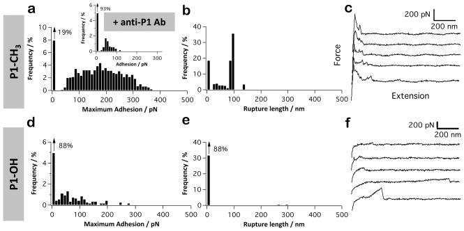
Figure 4.
P1 displays strong hydrophobic properties. (a–f) Adhesion force histogram (a and d), rupture length histogram (b and e), and typical force curves (c and f) obtained by recording force curves (n = 1024 curves for each condition) in PBS buffer with 1 mM Ca2+ between AFM tips functionalized with full-length P1 and methyl-terminated substrates (a–c) or hydroxyl-terminated substrates (d–f). The inset in (a) shows the adhesion force histogram obtained between P1 and a methyl-terminated substrate following blocking with a solution of monoclonal antibodies directed against the P1 head (mAb 1-6F). For each condition, similar data were obtained using at least 3 different tips and 3 different substrates.

