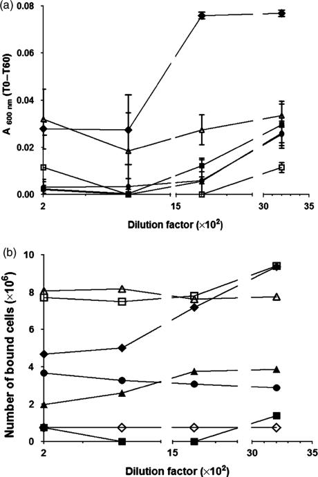
Fig. 6.
Functional characterization of antibodies raised in mice immunized with P139–512 produced in Bacillus subtilis. (a) Inhibition of saliva-mediated aggregation of Streptococcus mutans PC3370C cells. Samples containing bacterial cells and the tested serum were incubated for 5 min at 37 °C and the OD600nm was measured (T0). After an additional 60-min incubation period at 37 1C, the OD (T60) was measured once more. The values are expressed as the difference between T0 and T60 for each tested serum dilution. Symbols: (◆) nonimmune serum; (●) anti-P1 serum; (■) anti-P139–512 serum; (▲) anti-P139–512d serum; (□) anti-P1 serum adsorbed with P139–512; and (△) anti-P1 serum adsorbed with P139–512d. (b) Inhibition of S. mutans adherence to immobilized saliva in microplate wells. Streptococcus mutans PC3370C cells were mixed with diluted tested serum samples, incubated for 30 min to 37 °C, and transferred to microplate wells coated with clarified saliva and incubated for an additional 2 h. Bound cells were revealed by staining with 0.5% crystal violet and determination of absorbance of the solubilized cell-bound stain at 600 nm. Symbols are the same as those shown in (a) and (◇) cells incubated in the presence of 3 mM EDTA. All tested sera were diluted in order to reach a final titer of 104. Data represent the average of three independent experiments. Values are expressed as mean ± SD.

